Instrument
The WiscSIMS lab houses a CAMECA IMS-1280. The IMS-1280 is a large radius multi-collector ion microprobe incorporating many improvements over earlier instruments, several of which are designed to enhance precision and accuracy of isotope ratio analysis. The detector assembly includes a total of 10 electron multiplier and Faraday Cup detectors with five moveable trolleys for simultaneous analysis of a wide range of isotope systems. Both alkali metal (Cs+) and duoplasmatron (O-, O+) sources are available. Spot sizes as small as 250 nm are possible (with Cs), but more generally, a spot of 10 μm diameter by 1 μm deep is used to increase sample size (~1ng/analysis) and optimize accuracy and precision. Precision for δ18O and δ17O in well-polished silicates is typically ±0.3‰, (2 SD, spot-to-spot) with 10 μm diameter spots (Kita et al. 2009 Chem Geol; Valley & Kita 2009 MAC Shortcourse v 41).
A series of tutorials was presented by WiscSIMS personnel at the 2013 workshop “High Resolution Proxies of Paleoclimate”
- SIMS Basics (pdf; Noriko Kita, June 2013)
- Accuracy vs. Precision in SIMS Analysis (pdf; John Valley, June 2013)
- Analysis of Biogenic Carbonates (pdf; Reinhard Kozdon, June 2013)
- Approaches to Speleothem Analysis (pdf; Ian Orland, June 2013)
Instrument Modifications
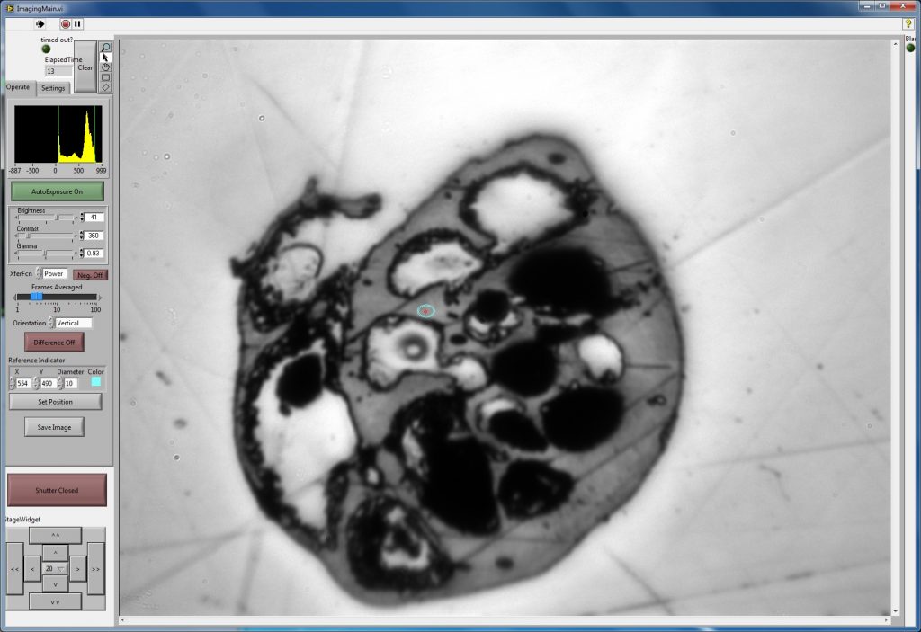
BadgerScope Software (2012)
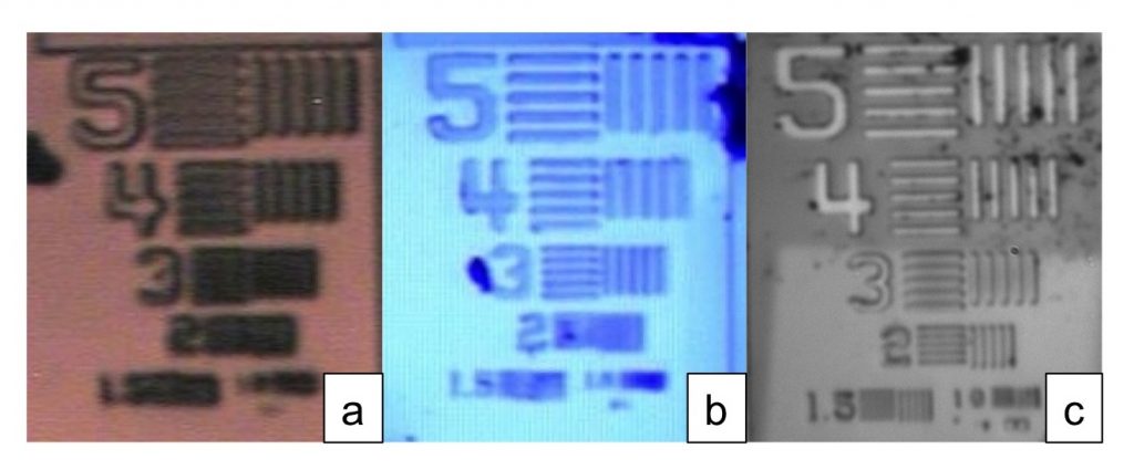
Reflected-light microscope images of a resolution-calibration target, CAMECA SIMS test sample. Numbers next to a set of lines (1.0 to 5) indicate width and distance between lines in micro-meters. Resolution of an image corresponds to the minimum width of lines that can be resolved. (a) Original CAMECA IMS 1280 microscope image with white light source. Resolution 3.5µm. Full color image. (b) Modification to blue LED light source (450nm). Resolution 2.5µm. Blue color image. (c) UV light system using LED light source (365nm). Resolution 1.3µm. Monochrome image.
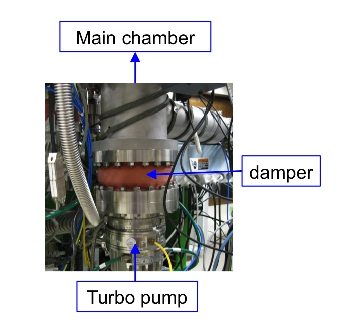
Different components of the machine.
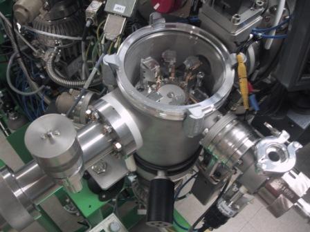
To increase pumping efficiency and ease sample transfer, we upgraded to a new CAMECA 6-sample airlock system in 2009.
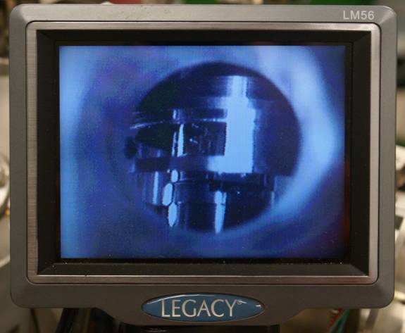
The CAMECA IMS 1280 ion probe has a sample arm that the user can manipulate to insert or remove samples from the sample stage. There is a small window positioned to allow the user to see the sample holder slide into the retaining clips on the sample stage. New users often had difficulties because the window is small, the chamber is unlit, and experience is needed to know where to look. To simplify viewing we’ve added an inexpensive camera, monitor, and power supply.
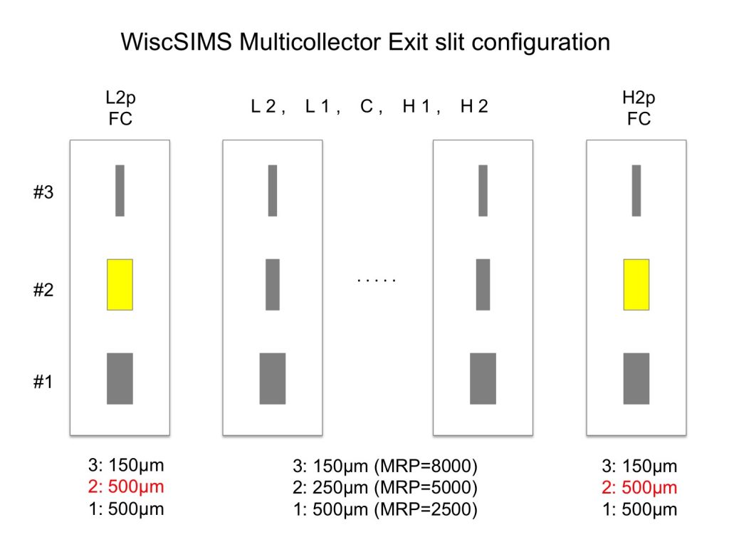
Mass resolution secondary ions using IMS 1280 Multicollection system is determined by the choice of three exit slit positions that are designed to move together for all detectors. When the instrument was installed in WiscSIMS lab, widths of three slits were made to be 500µm, 250µm, and 150µm, which correspond to Mass Resolving Power of 2500, 5000, and 8000 (assuming zero entrance slit width), respectively. Multicollectors are on the linear trolley that is not exactly along the focal plane of the mass spectrometer. Therefore, ions that are detected at the positions far from optical axis would be defocused at the exit slit plane. In the case of sulfur four (32, 33, 34, 36) isotope analyses, mass resolution of >4000 is required to separate 33S from 32SH interference by using slit #2 (250µm). However, by using 250µm exit slit width, 32S and 36S secondary ions do not show mass spectrum with wide flat top. For this reason, we replaced exit slit plates for two end detectors (L2p and H2p) by those having wider slit width for #2 (500µm). Mass resolution of 32S and 36S are set to ~2,200 with a wide flat top peak shape after the modification (Ushikubo et al. 2014) without any significant interference peaks.